image_banner_white
TDP 2025 photo adjusted

Thomas D. Pollard, MD

Sterling Professor Emeritus of Molecular Cellular and Developmental Biology, of Cell Biology and of Molecular Biophysics and Biochemistry, Yale University

Research Associate, Department of Molecular and Cell Biology

University of California, Berkeley

Personal 

Career summary (see CV for more information)

I retired from the full time Yale faculty in July 2021 and moved to Berkeley, CA, where I continue to do research at home and in the laboratory of David Drubin at UC Berkeley. Bill Earnshaw and I have authored of four editions of the textbook “Cell Biology” with illustrations by Graham Johnson.

In addition to research and teaching, I chaired my departments at the Johns Hopkins Medical School and Yale University, served as President of the Salk Institute for Biological Studies and was Dean of the Graduate School of Arts and Sciences at Yale. I served as president of both the American Society for Cell Biology and the Biophysical Society and in leadership positions at the National Academy of Sciences. For more details, see an autobiography at https://doi.org/10.1146/annurev-cellbio-100818-125427.

My honors include the Gairdner International Award, E.B. Wilson Medal from the American Society for Cell Biology, the Medal of Science from the State of Connecticut and election to the American Academy of Arts and Sciences, National Academy of Sciences and National Academy of Medicine.

Research summary (for more information see page on Research)

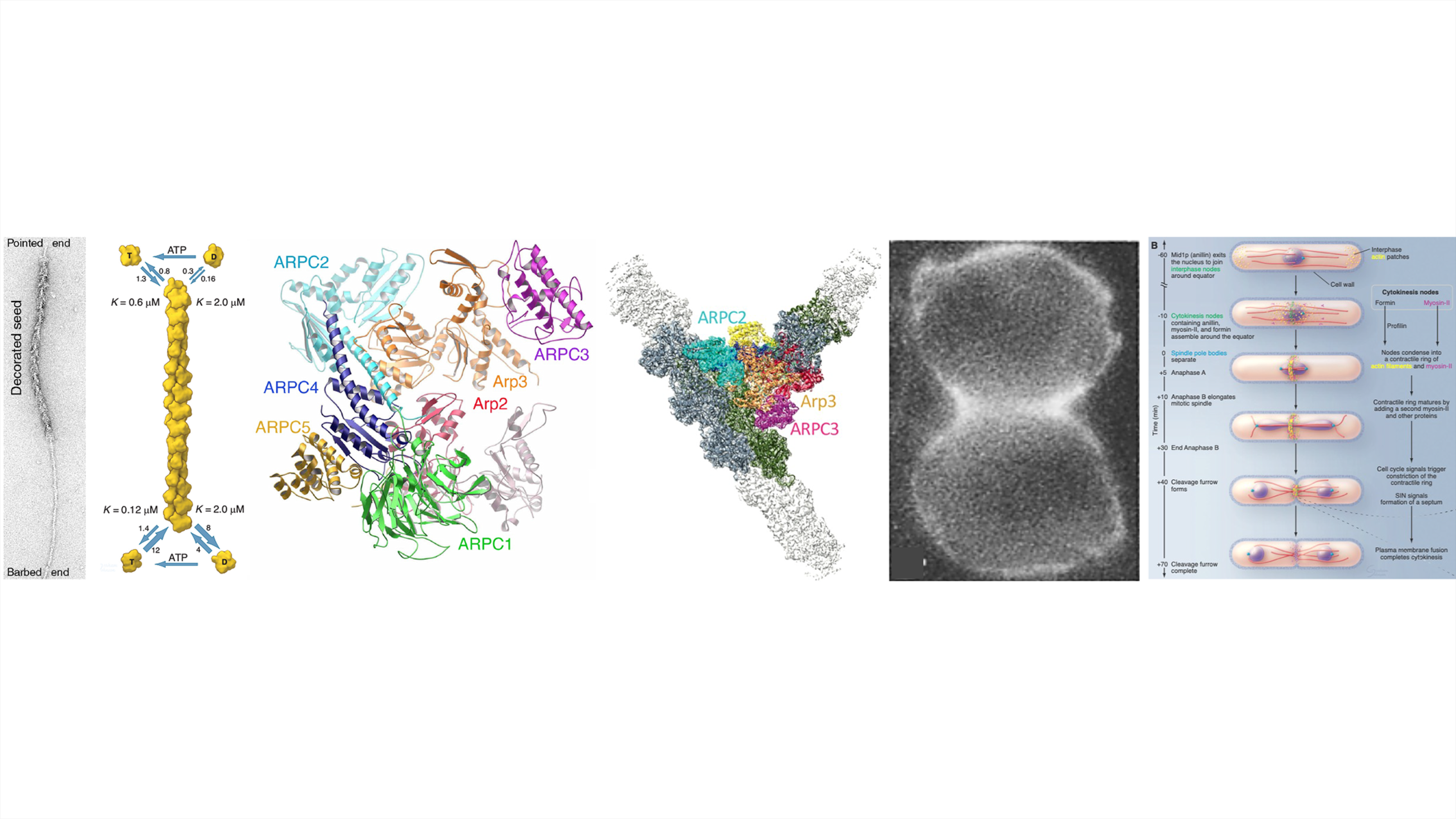
Starting in the 1960s, I have investigated along with members of my laboratory the molecular basis of cellular movements and cytokinesis using a combination of biochemistry, biophysics, microscopy and computational modeling.

Actin-based cellular movements are essential for shaping organs during embryonic development, defense against microorganisms and wiring the nervous system. Movement of cells out of primary tumors is the chief cause of mortality in cancer. Cytokinesis is essential for the replication of all cells and proliferation of cancer cells. 

My laboratory discovered and characterized many proteins that produce forces for cells to move including the first unconventional myosin (myosin-I), Arp2/3 complex and capping protein, all originally isolated from Acanthamoeba. We combined microscopy, biochemistry, biophysics, molecular genetics and mathematical modeling to provide the quantitative evidence required to formulate and test a detailed molecular explanation for how Arp2/3 complex stimulates the assembly of branched actin filaments that produce forces for cellular movements and endocytosis. We combined quantitative measurements of the time course of the appearance and disappearance of the participating proteins at site of endocytosis in fission yeast with mathematical modeling to confirm the molecular details and physics of the force-producing process.

Postdoc Keigi Fujiwara discovered myosin-II in the cleavage furrow of Hela cells in 1976, initiating our research on cytokinesis. In the late 1990’s the lab switched to using fission yeast to investigate cytokinesis. We characterized the participating proteins (actin, two isoforms of myosin-II, formin Cdc12, IQGAP Rng2, anillin Mid1p, F-BAR Cdc15, profilin, cofilin and capping protein) and measured the numbers of these proteins over time in the cytokinesis structures of live cells. This work culminated in the first molecularly explicit mathematical models and computer simulations of the mechanisms that assemble and constrict the cytokinetic contractile ring. 

Lab member news

Tom Pollard: In May 2025, I received the Connecticut Medal of Science, a biennial award from the State of Connecticut. This award recognizes the value of the work carried out in my laboratory by postdocs, graduate students, research assistants and collaborators over more than five decades. On this occasion, the Connecticut Academy of Science and Engineering interviewed me.

The Molecular Basis of Cellular Motility and Cytokinesis Interview

Medal of Science

Keigi Fujiwara: Keigi has been selected as Fellow of the American Society for Cell Biology. As a postdoc in the lab in the 1970s, he  used fluorescent antibodies to discover myosin-II in the cytokinetic contractile ring of HeLa cells. He is a retired Professor at MD Anderson Cancer Center, 

Fujiwara 2025 adjusted

Laura Machesky: Laura is President of the British Society for Cell Biology and Sir William Dunn Professor of Biochemistry at the University of Cambridge. As a PhD student in the 1990s, Laura discovered Arp2/3 complex and did seminal work on profilin. She has previously held faculty positions at University Birmingham and the Beatson Institute for Cancer Research.

Laura Machesky

Valda Vinson: Valda is Executive Editor for the Science family of journals. She manages the research content and editorial staff of five Science journals. As a postdoc in the 1990s Valda determined the first atomic structure of profilin by NMR and with Enrique De La Cruz carried out the definitive characterization of the interactions of profilin with ATP-, ADP- and nucleotide-free actin monomers, explaining how profilin catalyzes the exchange of ADP for ATP.

Valda Vinson

Michael Ostap: Mike is now Senior Vice Dean and Chief Scientific Officer for the Perelman School of Medicine at the University of Pennsylvania, in addition to maintaining his research laboratory as Professor of Physiology. As a postdoc in the 1990s, Mike was the first to establish the kinetic pathway of the enzyme cycle of an unconventional myosin, Acanthamoeba myosin-I.

Mike Ostap

Enrique De La Cruz: Enrique has been elected President of the Biophysical Society. His term starts in 2027. As a PhD student in the 1990s, he did classic work on the binding of ATP, phalloidin and profilin to actin monomers and filaments. He and Valda Vinson, explained how profilin catalyzes the exchange of ADP for ATP. He is now Professor of Molecular Biophysics and Biochemistry and Head of Branford College at Yale University.

EDLC small

Recent research news

I collaborate with members of Greg Voth’s laboratory at the University of Chicago using molecular dynamics simulations to characterize the structure and functions of actin filaments. Here are short summaries of the findings in recent papers.

Mechanism of phosphate release from actin filaments

• Wang Y, Wu J, Zsolnay V, Pollard TD, Voth GA (2024) Mechanism of Phosphate Release from Actin Filaments. Proc. Nat. Acad. Sci. USA. 121:e2408156121. PMID: 38980907. Molecular dynamics simulations revealed that the rate limiting step for the very slow release of phosphate from ADP-Pi-actin filaments is dissociation of phosphate from Mg2+ in the active site. A network of rapidly fluctuating hydrogen bonds in the release channel and a back door gate are not rate limiting.

Mechanism of Arp2/3 complex activation

• Iyer SS, WuJ, Pollard TD, Voth GA (2025) Molecular mechanism of Arp2/3 activation by nucleation promoting factors and actin monomer. Proc. Nat. Acad. Sci. USA. 122:e2421467122. PMID: 40048273. We used molecular dynamics simulations to estimate the rate and equilibrium constants for the activation of Arp2/3 complex by nucleation promoting factors prior to binding a mother filament and nucleating a daughter filament. Bound nucleation promoting factors increase the rate of the slow conformational change from inactive the “short-pitch” ten-fold. Binding to a “mother filament” stabilizes the active conformation and allows nucleation of a daughter filament.

Iyer image Arp2/3 complex
Actin filament structure at physiological temperature

• Iyer SS, Herman KM, Paul T, Wang I, Pollard TD, Voth GA (2025) Reaching the full potential of cryo-EM reconstructions with molecular dynamics simulations at 310 K: Actin filaments as an example. Proc. Nat. Acad. Sci. USA. 122:e2521421122. Molecular dynamics simulations at physiological temperature of cryo-EM structures of actin filaments revealed that freezing captures low entropy conformations, while higher entropy conformations predominate at physiological temperature. Thermal fluctuations of the filaments at 310 K explain the very slow rates of ATP hydrolysis and binding of cofilin.